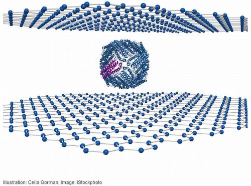
Biomolecule when sandwiched between sheets of graphene results in a high resolution of atomic level images of the molecule, a rare research put forth by the researchers at the University of Illinois, Chicago. The molecule called ferritin, an iron-storage protein, was imaged in the research.
Prior to this method, imaging of any Biomolecule was taken of course with an electron microscope along with a liquid stage container that is placed between thick windows of silicon nitrate to protect the sample from vacuum.
Graphene’s superior transparency becomes instrumental in providing an enhanced resolution relatively. And along with it, the sandwich accounts for the sample’s better protection from the electron beam that is fired during microscopy.
In order to avoid the deleterious effect of the electron beam, most of the electron microscopes prefer low energy beams, which further accounts for blurry images. However, the new research looks into the problem directly; extremely high thermal conductivity of graphene helps in combating the heat generated by the electron beam and similarly its property of high electrical conductivity helps in eradicating the heat of the shaft as it passes through the sample. Therefore, higher the energy beam would now correspond to higher resolution images unlike before.
Klie, the senior investigator on the research puts in,
the graphene sandwich will now open up analysis of biological and other difficult to image samples to almost anyone with an electron microscope.
Researchers envision that with this new form of technique involving graphene sandwich, preparation of samples could be done quickly and cheaply. Since the conventional liquid stage required huge investment in equipment and was an expensive preparation per sample.