Credits: UC San Diego News Center
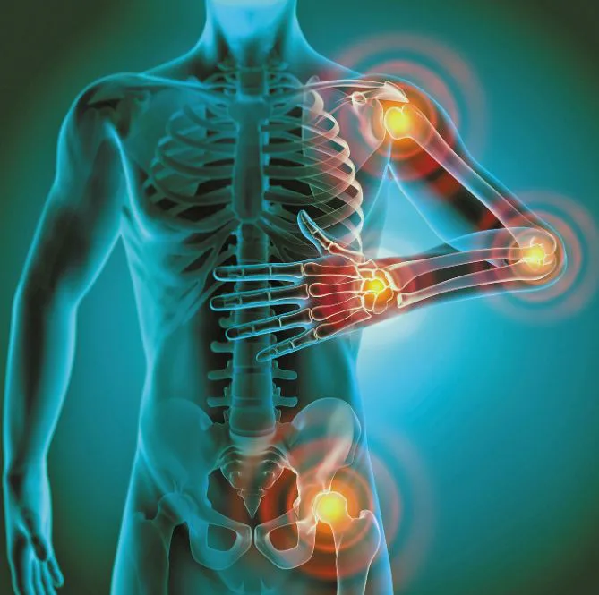
A spinal cord injury (SCI) usually causes perpetual damage within our body. This can result in long-term disability. And in most of the cases, spinal cord compression can lead to paralysis.
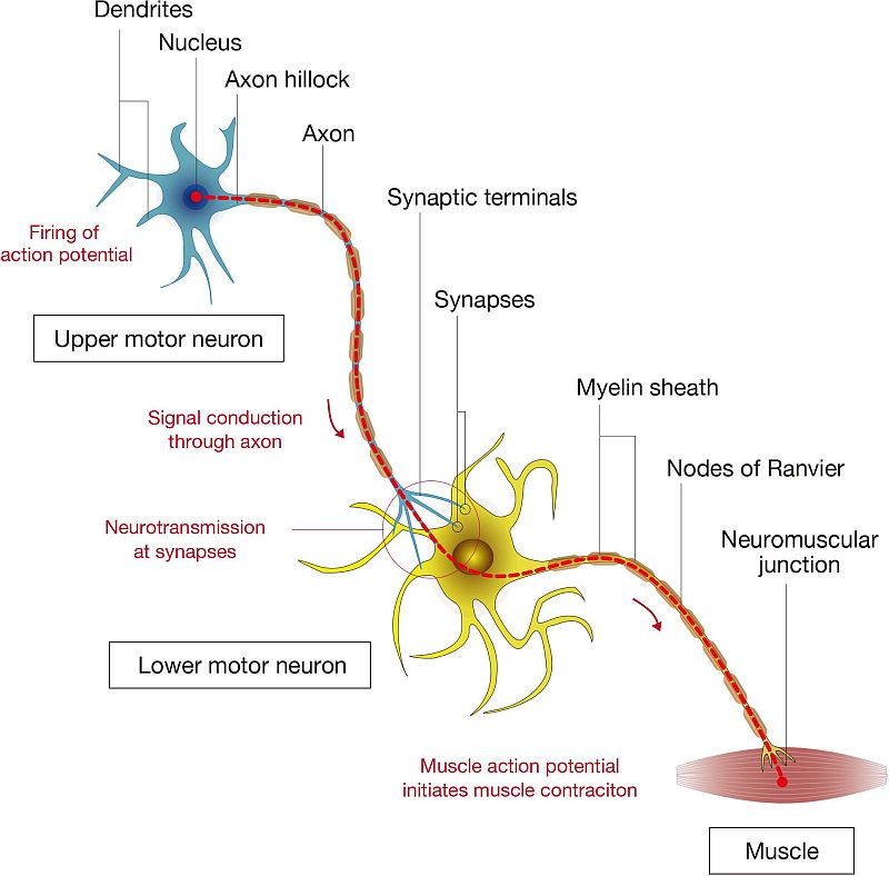
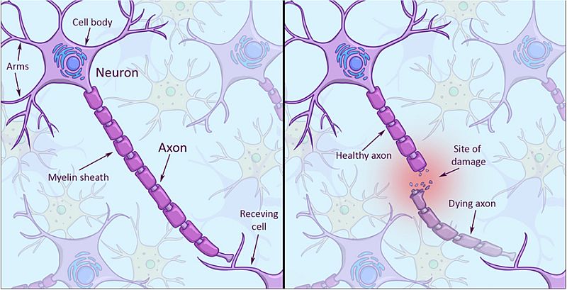
Damage to the spinal cord is a lifelong catastrophe. SCI rarely heals as injured nerve cells fail to regrow. The revival of nerve fibers, also called the slender projection of a nerve cell, is hindered by two things:
- scar tissue: although a scar helps nerve in attaching to a nearby tissue structure but it can also reduce the nerve’s blood supply.
- molecular processes inside the nerves: special molecules are present in neurons which helps in its growth. And these molecules are present only when neurons are active. Therefore, when an injury occurs, injured neurons are not able to process these molecules.
Not all nerves are irrecoverable. For instance, nerve cells present locations like the limbs, torso and nose have an ability to regrow and recuperate some or all of their function. However, neurons in the brain and spinal cord cannot regenerate.
Other signs and symptoms caused by damage to the nerve fibres in spinal cord are:
- Inability to move some or all of the body
- Intense stinging sensation, a prickly pain. As if the peripheral areas are injected with pins and needles.
- Loss or impaired sensation. A sense of numbness prevails where one is not able to feel heat, cold and touch
- Loss of bowel or bladder control
- Exaggerated reflexes or spasms. Reflex activity like, a knee-jerk reflex is stronger or amplified. Such a situation interferes with walking, movement and speech to name a few. While spasticity refers to stiffness or rigid muscles. In case of an internal injury, the muscles can lead to unusual tightness.
- Alteration in sexual health, that is, sexual sensitivity and fertility
- Trouble while inhaling and exhaling, including coughing or clearing secretions from lungs
Employing stems cells to resurrect the lost functions due to spinal cord injury has been one of the ambitious projects of neuroscientists so far. As per the World Health Organization, globally, 250 000 to 500 000 people suffer a spinal cord injury, yearly.
Credits: Frontiers in Cellular Neuroscience
Neural stem cell grafts
Recent study led by researchers at University of California San Diego School of Medicine has been a breakthrough in restoring lost functions due to SCI. Neuroscientists were able to implant extremely specialized grafts of neural stem cells directly into spinal cord injuries in mice.
Followed by documenting the subsequent growth and structural formation filling the injury sites.
Furthermore, the graft successfully latticed and mimicked the animals’ existing neural network.
So far, process till grafting was done however, the internal workings after integration was not quite known. No documentation was done before.
A previous research (on SCI in Monkeys) had shown signs of improved functioning in spinal cord injuries after neural stem cell grafts, however, neuroscientists did not know exactly what was happening.
Advantage of recent technological advances
Ceto one of the researchers said that they knew damaged host axons were expanding. To counteract that, the graft neurons were producing large number of axons into the spinal cord. But they were unable to record any activity taking place inside the graft site.
Ceto and the team leveraged the advanced technological options that they had. And so, they were able to simulate and track
- the developmental activity of the graft since it is latticed into the injury site and
- the structural orientation of neuron populations with light rather than electricity
This confirmed exactly which host and graft neurons were in play. What activities are being done across the tissue than recording any misleading results.
They concluded that graft neurons responded robustly. They were able to fire naturally in distinct clusters similarly like the neural networks of the normal spinal cord.
Graft neurons were also able to process sensory stimuli, like light touch and pinch.
Credits: Frontiers for Young Minds
Neural stem cell grafts are functional
Ceto and team collected all the results and claimed that not only the neural stem cell grafts have a tendency to self-assemble into (natural) neural networks much like spinal cord. But it can also functionally assimilate with the host nervous system. A speculation that has been finally proved.
Researchers’ next step is to organize grafts at topological level so that they can copy normal spinal cord with scaffolds and using electrical stimulation. In order to create robust synapses between host and graft neurons using electrical stimulation.
They envision to a future where stem cells, stimulation, rehabilitation and other technologically advanced interventions will together converge to develop therapy for patients dealing with spinal cord injury.
Via: Scott LaFee, University of California, San Diego